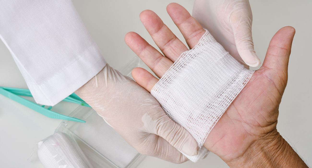
The journey following a surgical procedure often shifts focus entirely onto the healing of the incision, an intricate biological process that moves beyond the skill of the surgeon and into the complex, autonomous mechanisms of the body. A surgical wound, unlike an accidental trauma, is a planned, clean injury, meticulously created to allow access to underlying tissues. Understanding the systematic progression of its repair is essential for both the patient and the clinician, as deviations from the expected sequence are the first indicators of a complication that requires intervention. This process is not a smooth, linear progression but a cascade of overlapping biochemical and cellular events, each stage dependent on the successful completion of the one preceding it, a biological choreography where success relies on a delicate, almost improbable balance of forces.
A surgical wound, unlike an accidental trauma, is a planned, clean injury, meticulously created to allow access to underlying tissues
The body’s immediate, almost instantaneous response to the sharp incision is the phase known as haemostasis, a critical survival mechanism. This process is triggered by the disruption of blood vessels, prompting an immediate and transient vasoconstriction to minimize blood loss. Following this initial reflex, platelets swiftly adhere to the exposed collagen fibers at the site of injury, aggregating to form a temporary platelet plug. These activated platelets then release a complex array of chemical mediators and growth factors that initiate the coagulation cascade. Fibrinogen is converted into a fibrin mesh, which reinforces the platelet plug, creating a stable, resilient clot that effectively seals the compromised vasculature. This entire phase, while brief, is foundational; a failure here can result in hematoma formation—a collection of blood that separates tissue layers—which itself can become a fertile ground for bacterial proliferation and ultimately delay the entire healing trajectory.
Following this initial reflex, platelets swiftly adhere to the exposed collagen fibers at the site of injury, aggregating to form a temporary platelet plug
Almost immediately overlapping with the final stages of coagulation is the inflammatory phase, which is less a pathology and more a necessary preparatory step for repair. It is marked by the classic clinical signs of erythema, heat, edema, and tenderness, which are direct consequences of increased vascular permeability and localized vasodilation. The purpose is defensive: neutrophils, the first wave of immune cells, infiltrate the wound space to scavenge bacteria and debris, setting the stage for macrophages. These macrophages arrive slightly later, taking on the indispensable role of clearing cellular waste, broken down matrix components, and effete neutrophils. Beyond their housekeeping duties, macrophages are the primary orchestrators of the transition to the next stage, releasing an array of powerful growth factors and cytokines that signal the commencement of constructive tissue rebuilding. If this inflammation persists beyond a few days, it becomes a sign that the wound is struggling, indicating a potential chronic issue or an underlying pathogenic colonization.
Beyond their housekeeping duties, macrophages are the primary orchestrators of the transition to the next stage, releasing an array of powerful growth factors and cytokines
The proliferative phase, beginning approximately two to four days post-incision, is fundamentally about reconstruction. The visible evidence of this stage is the development of granulation tissue, a soft, red, and moist matrix that fills the wound defect. This tissue is rich in fibroblasts, cells that begin to synthesize and deposit new collagen, a key structural protein that lends tensile strength to the developing scar. Simultaneously, angiogenesis is rampant, with new capillary networks budding from existing vessels to provide the necessary oxygen and nutrients to the metabolically active wound bed. At the wound edges, epithelial cells begin to migrate and multiply, creating a protective barrier across the surface of the injury in a process known as epithelialization. The wound begins to visibly contract as specialized fibroblasts pull the edges inward, reducing the overall size of the defect. Any interruption to this delicate balance—such as desiccation or mechanical tension—can significantly impair the formation of this essential, temporary scaffolding.
This tissue is rich in fibroblasts, cells that begin to synthesize and deposit new collagen, a key structural protein that lends tensile strength to the developing scar
The final and longest stage is remodeling, or maturation, a phase that can extend from several weeks to over a year after the incision has physically closed. During this time, the hastily deposited, disorganized Type III collagen is gradually broken down and replaced by the stronger, more robust Type I collagen. The collagen fibers are constantly reorganized, cross-linked, and aligned along the lines of mechanical stress to improve the strength and structure of the scar. While the healed wound will never regain the original tensile strength of the undamaged skin, its strength significantly increases during this period. The scar tissue also begins to lose its initial raised, red appearance as vascularity decreases and the cellular activity subsides, transitioning to a flatter, paler, and less conspicuous mark. It is in this phase that the patient’s long-term care, including gentle massage and protection from sun exposure, can influence the final cosmetic outcome of the scar.
While the healed wound will never regain the original tensile strength of the undamaged skin, its strength significantly increases during this period
Surgical wounds are typically closed by primary intention, meaning the surgeon meticulously approximates the edges using sutures, staples, or adhesive materials, which facilitates the most rapid epithelialization and minimizes scar formation. However, certain clinical situations preclude this direct closure. Wounds that are highly contaminated, those with significant tissue loss, or those at high risk of infection may be left open to heal by secondary intention. In secondary healing, the wound must close entirely through the formation of granulation tissue, contraction, and subsequent epithelialization, a significantly slower process that inevitably results in a larger, more prominent scar. Less common is tertiary intention, or delayed primary closure, where a wound is left open initially for a period to manage contamination or swelling and is then surgically closed once the local conditions are deemed favorable. The choice of closure technique has profound implications for both the management protocol and the patient’s recovery timeline.
Wounds that are highly contaminated, those with significant tissue loss, or those at high risk of infection may be left open to heal by secondary intention
The success of these healing stages is continuously modulated by an intricate web of intrinsic and extrinsic factors that operate both locally and systemically. Intrinsic factors include the patient’s general health status: conditions like poorly controlled diabetes, which impairs immune function and circulation; chronic diseases that lead to poor tissue oxygenation (hypoxia); and age-related decreases in tissue elasticity and immune response. Extrinsic factors often relate to surgical technique and post-operative management, such as the presence of foreign material (sutures or drains), wound tension, or the formation of an internal fluid collection like a seroma. Furthermore, lifestyle choices like smoking, which severely compromises tissue oxygen delivery, and malnutrition, which starves the body of the necessary protein and vitamin building blocks, can profoundly impede the orderly progress of repair, diverting an acute wound onto the pathway toward chronicity.
Furthermore, lifestyle choices like smoking, which severely compromises tissue oxygen delivery, and malnutrition, which starves the body of the necessary protein and vitamin building blocks, can profoundly impede the orderly progress of repair
Recognizing the subtle yet critical signs of a healing complication is paramount for effective intervention. While a degree of redness, mild swelling, and tenderness is an expected part of the necessary inflammatory process during the first few days, signs that are escalating in severity, spreading beyond the wound margin, or persisting past the expected timeframe are red flags. The appearance of purulent discharge, increasing pain, a foul odor, or fever are clear clinical markers of a surgical site infection. A more mechanically focused complication is dehiscence, where a wound previously closed by primary intention opens up, potentially exposing deeper tissues. Any sign of a wound that is not visibly improving after the first week, or one that exhibits excessive, dark-colored granulation tissue, warrants immediate clinical re-assessment, as the window for effective, non-invasive treatment can be narrow.
The appearance of purulent discharge, increasing pain, a foul odor, or fever are clear clinical markers of a surgical site infection
Contemporary wound care moves far beyond simple gauze and tape, incorporating a spectrum of advanced therapeutic modalities to address complex or impaired surgical sites. Negative Pressure Wound Therapy (NPWT), for instance, utilizes a vacuum to continuously draw exudate and infectious material from the wound bed, reducing edema, promoting blood flow, and encouraging the formation of healthy granulation tissue. Specialized dressings, such as alginates and hydrogels, are used to manage different wound environments, with some absorbing excessive fluid while others maintain a critical level of moisture necessary for cell migration and tissue development. For non-healing chronic wounds, advanced biological approaches, including the use of skin substitutes or the local application of platelet-rich plasma, may be employed to deliver essential growth factors and cellular components directly into the compromised tissue. These targeted interventions are crucial in rescuing stalled healing processes and preventing catastrophic tissue loss.
Negative Pressure Wound Therapy (NPWT), for instance, utilizes a vacuum to continuously draw exudate and infectious material from the wound bed, reducing edema, promoting blood flow, and encouraging the formation of healthy granulation tissue
The recovery from a surgical procedure is truly a testament to the body’s innate capacity for self-repair, a complex, staged endeavor defined by the precise and dynamic interplay of cellular signaling and matrix synthesis. It is a process that can be derailed by systemic vulnerabilities and local complications, requiring careful and continuous clinical oversight. Success is ultimately measured not only by the functional return of the operated site but also by the quality of the healed tissue, a visible, tangible reminder of a complex biological triumph over injury.
